Application of live cell imaging in the study of central nervous system (CNS) diseases and disorders
Studying the causes of human central nervous system (CNS) diseases and disorders in order to find effective treatments requires in vitro and in vivo disease models that truly reproduce their neuropathophysiological conditions and also support neurons through the necessary cellular mechanisms. Responses to the treatments that provide translation results 1-3 .
In addition, we need to study the earliest neuropathological changes, as these changes provide an excellent opportunity for early diagnosis and intervention. Structural and functional changes in synapses are often the earliest pathological changes in many neurodegenerative and central nervous system diseases.
To detect early synaptic dysfunction, scientists used live cell imaging to provide single-cell images and analyze cell morphology changes in real-time as experimental conditions change. This communication discusses the use of live cell imaging in in vitro and in vivo studies of central nervous system diseases and diseases.
Designing biologically relevant in vitro and in vivo live cell imaging experiments requires healthy, mature neurons that require appropriate synaptic structures and functions (ie, morphology and electrical activity) (below). The primary imaging targets are presynaptic and postsynaptic structures and related proteins (eg, synaptophysin, neurotransmitter receptors, ion channels, scaffold proteins or cytoskeletal proteins [F-actin, microtubules]) because They are the first substrates to undergo pathological changes 4 (below).
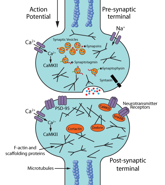
Methods for assessing changes in synaptic structure or function include measuring synaptic density, axonal length, spinal density, nodes (position of axons on the cell body), dendritic branches, or electrical activity 4 .
Live cell imaging of mammalian neurons cultured in vitro, including human-derived pluripotent stem cell (hipsc)-derived neurons, due to its greater biological relevance to human neurons 8 , can reveal neural regression with humans Unique mechanisms and functional discoveries associated with sexual diseases such as Alzheimer's disease (AD) and Parkinson's disease. Apolipoprotein E (apoe-E) is a protein closely related to the pathogenesis of AD, but the role of apoe-E and its metabolites in neuronal physiology has not been taken seriously.
Recent studies have found, differentiated SH-SY5Y neuroblastoma exhibits neural hyperplasia (assessed by live cell imaging and neurite length positive changes in cell fusion) After 9 full-length 25kDa apoe-E fragment, or long-term treatment, the initial drug screening Research has also benefited from this approach. In AD, the two most common drug targets are amyloid beta (aβ) and tau, both of which have early pathological effects on synapses 5-7 .
In recent years, some scholars have performed live cell imaging of HIPSC-derived neurons, and screened several candidate antibodies against pathological Aβ, and quantified axonal length and dendritic branch points by incubating time and concentration range of different gradient antibodies. Changes to assess positive immunotherapy outcomes 10 .
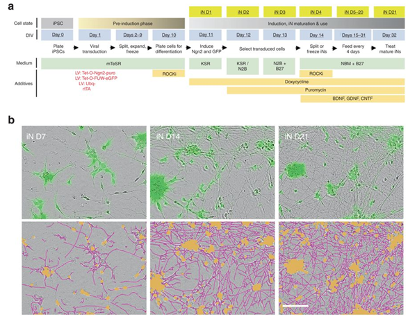
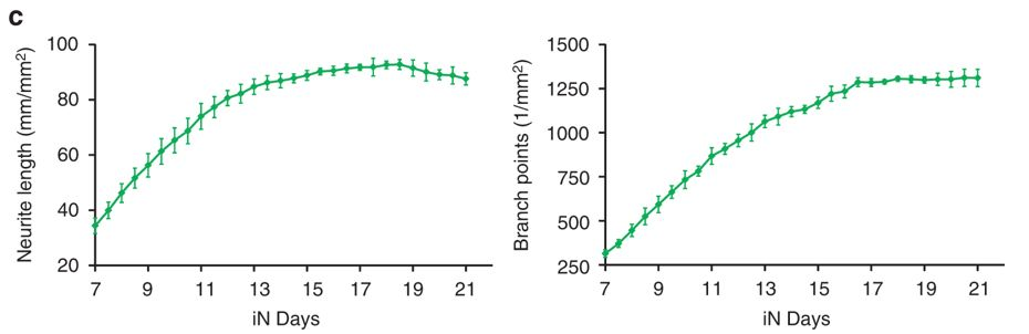
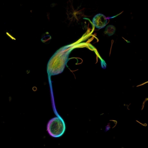
Live cell imaging of 0-21 days of human induced neuron (INS) differentiation
In another study, Aβ, Hong et al HIPSC 11 pairs of a living body-derived neurons were imaged using isolated from postmortem human brain extracts Aβ oligomers of these neurons processed by quantitative analysis of axonal Changes in length and synaptic plasticity, combined with long-term potential recording, were used to assess the pathophysiological properties of the oligomer.
With the birth of the first resonance-enhanced resonance energy transfer (FRET) sensor, which is sensitive to the tau conformation, tau protein has become the focus of a new technology that monitors live hela cells and immortalized HT22 hippocampal neurons. After drug treatment, the conformation of wild-type and pathologically mutated tau in the presence and absence of microtubules (mts) 12 , this tau-FRET sensor provides invaluable quantitative and qualitative data on the regulation of tau binding to mts The ratio of soluble to insoluble (pathological) tau and how it affects the conformation of tau to facilitate non-pathological forms.
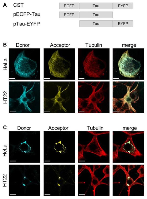
Imaging of Tau Protein Conformation Sensitive Sensor (CST) in Living Cells
In vivo animal models are complementary to in vitro cell culture models, and live cell imaging of animal neurons is typically performed using a two-photon microscope that allows observation of functional neurons in a natural state and in a larger environment of interconnected support networks. Cell Image 13 In this way, live cell imaging in animals is revolutionary, and researchers have the opportunity to visually analyze cellular dynamics in real time.
In addition, a single cell in vivo imaging may feeling, behavioral changes, and functional response of neurons during drug treatment quantify 13. In vivo two-photon live cell imaging is used to find the mechanism of action of α-synuclein targeting anti- Parkinson (PD) drugs 14 and to evaluate the transplantation of embryonic cells into damaged adult brain regions to replace death or imminent death. The feasibility and optimal timing, host and donor conditions of neuron 15-17 .
In addition, in vivo imaging can reveal new therapeutic approaches. Live cell imaging of MPTP mouse PD model shows that the structural and functional synaptic plasticity of MPTP-treated mouse motor cortical neurons is affected by the selective loss of dopaminergic neurons, which improves regulation. Neuronal activity in specific areas of the motor cortex is a viable option for treating PD-related sports injuries 18 .
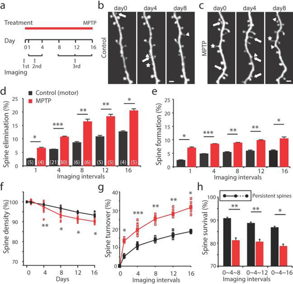
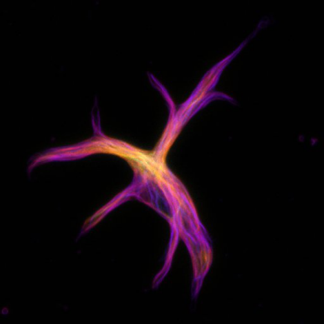
Dynamic changes of the motor cortex in the PD mouse model
to sum up
In vivo cell imaging is a valuable tool for studying diseases and disorders of the central nervous system. It not only allows researchers to gain an in-depth understanding of basic disease/disorder biology, but also provides real-time insight into the functional consequences of pharmacological, genetic, and behavioral therapeutic interventions. Coupled with advances in super-resolution microscopy and automated live cell imaging 19 , we have a good opportunity to focus on the first pathological synaptic changes that can be stopped or reversed by early therapeutic intervention.
Amytech can provide you with a full range of valuable research tools such as Cytoskeleton's live cell imaging probes for f-actin, microtubules, DNA and lysosomes, as well as purified cytoskeletal proteins, activation assays and Antibodies; iRegene's human neural stem cell (hNSC) cell line, supporting stem cell culture medium and neuron-directed medium; StressMarq's active alpha synaptic protein and Tau protein, etc., can greatly promote these studies. effect.
Cytoskeleton's live cell imaging related products:
Live Cell Imaging Kit:
| product name | Item number |
| SiR-Actin Kit | (Cat. # CY-SC001) |
| SiR-Tubulin Kit | (Cat. # CY-SC002) |
| Cytoskeleton Kit (including SiR-Actin, SiR-Tubulin, and Verapamil) | (Cat. # CY-SC006) |
| SiR-DNA Kit | (Cat. # CY-SC007) |
| SiR-Lysosome Kit | (Cat. # CY-SC012) |
| SiR700-Actin Kit | (Cat. # CY-SC013) |
| SiR700-Tubulin Kit | (Cat. # CY-SC014) |
| SiR700-DNA Kit | (Cat. # CY-SC015) |
| SiR700-Lysosome Kit | (Cat. # CY-SC016) |
Factory internal quality inspection:
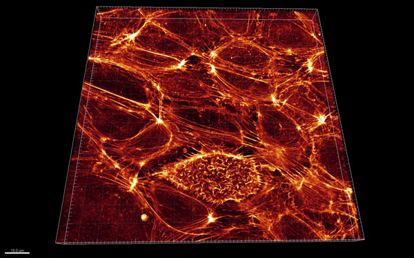 | Huvec cells: Â Huvec cells (fixed) were stained with SiR-actin and imaged by confocal microscopy. |
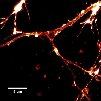 | Rat hippocampal neurons: STED images of rat hippocampal neurons cultured by SiR-actin staining. The actin ring (streak) is periodically 180 nm. |
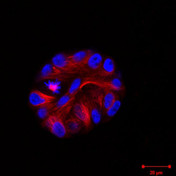 | MCF10A Cells (3D culture ) MCF10A cells stained with SiR-actin (red) expressed H2B-GFP (blue) in Matrigel. LSM inverted microscope observation chart |
 | MCF10A Cells (3D culture ) mf10a cells stained with SiR-actin (red) expressed H2B-GFP (blue) in Matrigel. LSM inverted microscope observation chart |
 | Goldfish Retinal Bipolar Cells: Three-dimensional projection of single isolated goldfish retinal bipolar cells stained with SiR-tubulin. The color spectrum represents depth. It can be seen that the microtubules protrude from the top (top) of the dendrite into the axon and extend downward into the giant synaptic end (bottom). |
 | Goldfish astrocytes: Three-dimensional projection of single isolated goldfish astrocytes stained with SiR-tubulin. |
G-LISA Activation Detection Kit:
| product name | Item number |
| RhoA G-LISA Activation Detection Biochemical Kit (Colorimetric Method) | (Cat. # BK124) |
| RhoA G-LISA Activation Detection Biochemical Kit (Fluorescence) | (Cat. # BK121) |
Actin Biochemical Kit:
| product name | Item number |
| Actin-binding protein Spin-Down biochemical assay kit (rabbit skeletal muscle actin) | (Cat. # BK001) |
| Actin-binding protein Spin-Down biochemical assay kit (human platelet actin) | (Cat. # BK013) |
| Actin Polymerization Biochemical Test Kit (Fluorescence: Rabbit Skeletal Actin) | (Cat. # BK003) |
| G-Actin/F-actin In Vivo Biochemical Test Kit | (Cat. # BK037) |
Tubulin Biochemical Kit:
| product name | Item number |
| Tubulin Polymerization Biochemical Test Kit (Colorimetric Method) | (Cat. # BK006P) |
| Tubulin Polymerization Biochemical Test Kit (Fluorescence) | (Cat. # BK011P) |
| Microtubule-binding protein Spin-Down biochemical detection kit | (Cat. # BK029) |
| Microtubule/Tubulin In Vivo Biochemical Test Kit | (Cat. # BK038) |
iRegene 's human stem cell line and supporting culture kit:
| product name | Item number |
| NouvNe uTM human neural stem cell (hNSC) cell line | (RJC02006) |
| NouvNeu!" hNSC Neural Stem Cell Culture Kit | (RJM02000) |
| NouvNeuTMhNeuron Neuron Oriented Differentiation Cell Culture Kit | (RJM03000) |
StressMarq 's active alpha synaptic protein and Tau protein facilitate the rapid construction of animal models of neurological diseases:
| product name | active | Application Type | Item number | Applicable species |
| Recombinant human α-synuclein monomer (control) recombinant human α-synuclein polymer PFFs (control)  | Inactive | WB, SDS-PAGE, in vivo/vitro | SPR-316B SPR-317B |  |
| Recombinant human α-synuclein monomer recombinant human α-synuclein polymer PFFs | Active | WB, SDS-PAGE, in vivo/vitro | SPR-321B SPR-322B |  |
| Recombinant mouse α-synuclein monomer recombinant mouse α-synuclein polymer PFFs | Active | WB, SDS-PAGE, in vivo/vitro | SPR-323B SPR-324B |  |
| Mouse anti-mouse α-synuclein monoclonal antibody mouse anti-mouse α-synuclein monoclonal antibody mouse anti-mouse α-synuclein monoclonal antibody |  | WB, ICC/IF WB, IHC, ICC/IF WB, IHC, ICC/IF | SMC-531DSMC-532DSMC-533D | Human, mouse, rat, mouse, rat, mouse, rat |
| Mouse anti-human α-synuclein monoclonal antibody against human α-synuclein polyclonal antibody |  | WB, ICC/IF WB | SMC-530DSPC-800D | Human, mouse, rat |
| Alpha-synuclein (phosphorylated Ser129) antibody alpha-synuclein (phospho-Tyr136) antibody | Â | WB, ICC/IF WB | SPC-742DSPC-1435D | Human, mouse, mouse, rat |
| Recombinant Tau 2N4R P301S protein monomer recombinant Tau 2N4R P301S protein PFFs | Active | WB, SDS-PAGE, in vivo/vitro | SPR-327B SPR-329B | Â |
| Recombinant Tau K18/P301L Protein Monomer Recombinant Tau K18/P301L Protein PFFs | Active | WB, SDS-PAGE, in vivo/vitro | SPR-328B SPR-330B |
references:
- Neurological disease models made clear. Med. (Editorial, published September 2015). 21 , 964.
- Schlachetzki JC et al. 2013. Studying neurodegenerative diseases in culture models. Bras. Psiquiatr. 35 , S92-100.
- Hughes P. et al. 2018. The costs of using unauthenticated, over-passaged cell lines: how much more data do we need? Biotechniques . 43 , 575, 577-578, 581-582
- Verstraelen P. et al. 2018. Image-based profiling of synaptic connectivity in primary neuronal cell culture. Neurosci . 12 , 389.
- Hoover BR et al. 2010. Tau mislocalization to dendritic spines mediates synaptic dysfunction independently of neurodegeneration. Neuron . 68 , 1067-1081
- Spires-Jones TL and Hyman BT 2014. The intersection of amyloid beta and tau at synapses in Alzheimer's disease. Neuron . 82 , 756-771.
- Bayer TA and Wirths O. 2010. Intracellular accumulation of amyloid-beta – a predictor for synaptic dysfunction and neuron loss in Alzheimer's disease. Front. Aging Neurosci . 2 , 8
- Unternaehrer JJ and Daley GQ 2011. Induced pluripotent stem cells for modelling human diseases. Trans. R. Soc. B. 366 , 2274-2285.
- Munoz SS et al. 2018. The serine protease HtrA1 contributes to the formation of an extracellular 25-kDa apolipoprotein E fragment that stimulates neuritogenesis. Biol. Chem . 293 , 4071-4084
- Jin M. et al. 2018. An in vitro paradigm to assess potential anti-Aβ antibodies for Alzheimer's disease. Commun. 9 , 2676.
- Hong W. et al. 2018. Diffusible, highly bioactive oligomers represent a critical minority of soluble Aβ in Alzheimer's disease brain. Acta Neuropathol. 136 , 10-40
- Di Primio C. et al. 2017. The distance between N and C termini of tau and of FTDP-17 mutants is modulated by microtubule interactions in living cells. Neurosci. 10 , 210.
- White MD et al. 2018. In vivo imaging of single mammalian cells in development and disease. Trends Mol. Med. 24 , 278-293.
- Wrasidlo W. et al. 2016. A de novo compound targeting alpha-synuclein improved deficits in models of Parkinson's disease. 139 , 3217-3236.
- Falkner S. et al. 2016. Transplanted embryonic neurons integrate into adult neocortical circuits. Nature . 539 , 248-253.
- Peron S. et al. 2017. A delay between motor cortex lesions and neuronal transplantation enhances graft integration and improves repair and recovery. Neurosci. 37 , 1820-1834.
- Wang C. et al. 2016. Infiltrating cells from host brain restore the microglial population in grafted cortical tissue. Rep. 6 , 33080.
- Guo L. et al. 2015. Dynamic rewiring of neural circuits in the motor cortex in mouse models of Parkinson's disease. Neurosci. 18 , 1299-1309.
- Shin HY et al. 2018. Using automated live cell imaging to reveal early changes during human motor neuron degeneration. eNeuro . doi: 10.1523/ENEURO.0001-18.2018.
As the regional distributor of Cytoskeleton, iRegene and StressMarq in China, Aimei Technology Co., Ltd. will provide Chinese customers with the most comprehensive Cytoskeleton, iRegene, StressMarq products and services.

Laxative Powder,Laxaday Powder,Smooth Lax Powder,Constipation Relief Powder
Shaanxi Zhongyi Kangjian Biotechnology Co.,Ltd , https://www.zhongyibiotech.com